AI in Medical Imaging: What Patients and Clinicians Should Know
Learn how artificial intelligence supports X-rays, CT, MRI, ultrasound and cancer screening, including its potential benefits, limitations, safety risks and FDA oversight.
At a glance
What AI can do in medical imaging | What it does not mean |
|---|---|
Reconstruct or improve images | That every scan can be performed faster or with less radiation |
Measure or segment anatomy | That the measurement is always correct |
Flag or prioritize suspected findings | That the software has made a complete diagnosis |
Support comparison with prior studies | That the clinical history is no longer needed |
Assist reporting and workflow | That radiologists or other qualified clinicians are no longer responsible for interpretation |
Artificial intelligence is now used across many parts of medical imaging, from image reconstruction and measurement to detection, triage and workflow support. The most important point for patients and clinicians is that these systems are designed for specific tasks. Their usefulness depends on what they were built to do, how well they were validated, the population and equipment in which they are used, and how their output is reviewed.
Medical imaging can generate hundreds or thousands of images from a single examination. A radiologist may need to interpret those images alongside symptoms, laboratory results, previous scans and the wider clinical history. AI can help with parts of that workload, but a highlighted region, probability score or automated measurement is not the same as a complete clinical diagnosis.
This 2026 update also reflects several newer developments: the FDA's expanding AI-enabled medical-device list, growing attention to trustworthy and explainable imaging AI, biomedical foundation models, multimodal systems and early work on agentic AI in radiology.
For a broader overview of clinical documentation, monitoring, research and hospital operations, see Artificial Intelligence in Healthcare: Uses, Benefits, Risks and Examples in 2026.
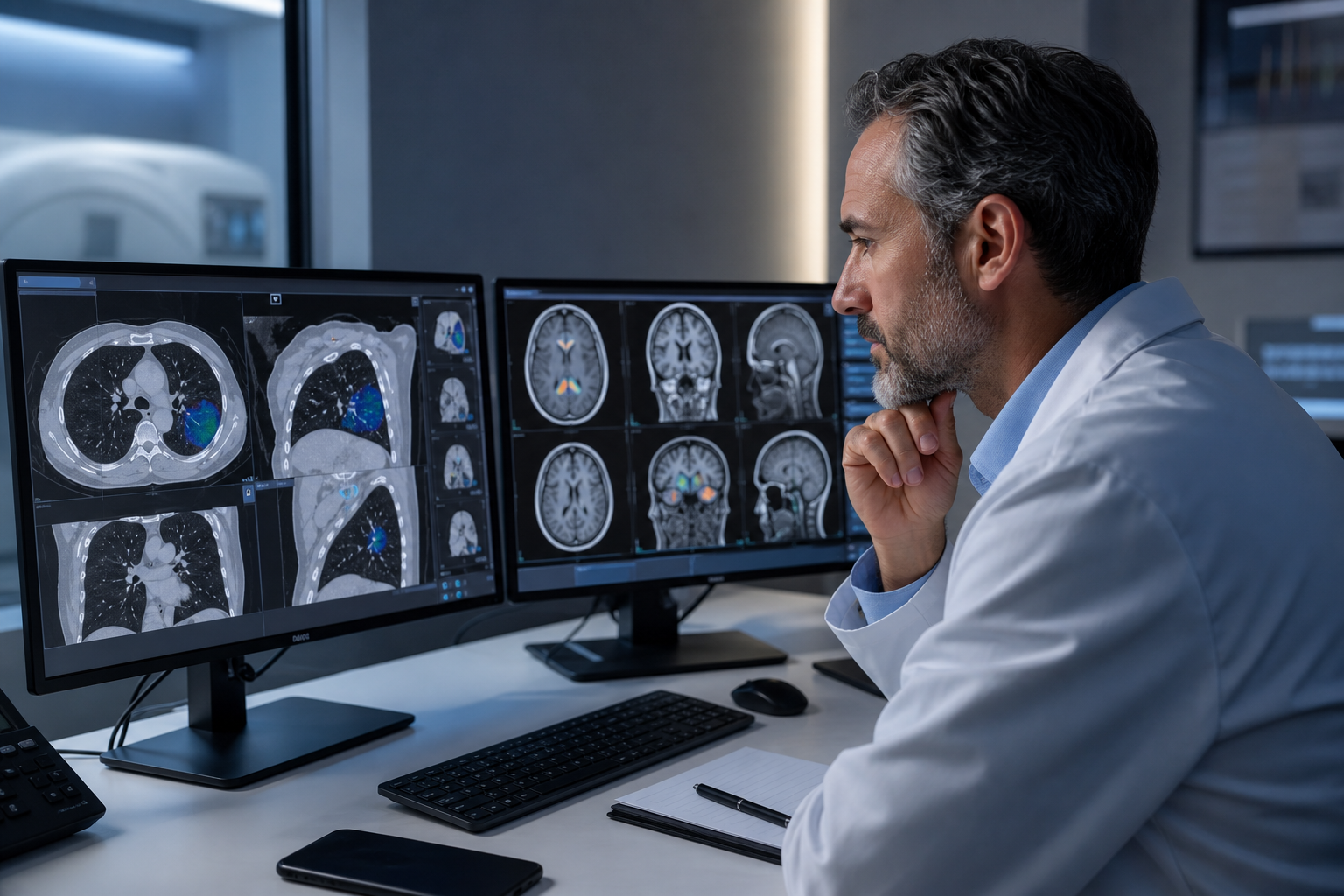
A radiologist reviews AI-assisted highlights alongside the original medical images. The software can draw attention to patterns, but the clinician remains responsible for interpretation. Credit: Biomed Atlas editorial illustration.
What is AI in medical imaging?
AI in medical imaging refers to software that uses computational models to perform a defined task involving images such as X-rays, mammograms, CT scans, MRI scans, ultrasound, nuclear medicine studies, retinal photographs or digital pathology images.
Depending on the system, AI may:
help reconstruct an image or reduce noise;
outline an organ, tumor or other structure;
measure size, volume, density or change over time;
flag a suspected abnormality for clinician review;
prioritize an examination that may contain an urgent finding;
compare a current study with prior imaging;
support treatment planning or response assessment;
or assist workflow tasks such as protocol selection and report preparation.
These functions should not be treated as interchangeable. A device authorized to identify a particular finding on a chest CT, for example, should not automatically be assumed to diagnose every possible abnormality on the scan.
How does AI-assisted imaging work?
A simplified workflow starts with image acquisition. The AI system then processes the images and returns the output it was designed to generate, such as an outline, measurement, alert, probability score or reconstructed image. A qualified clinician reviews that output together with the original examination and the patient's clinical information.
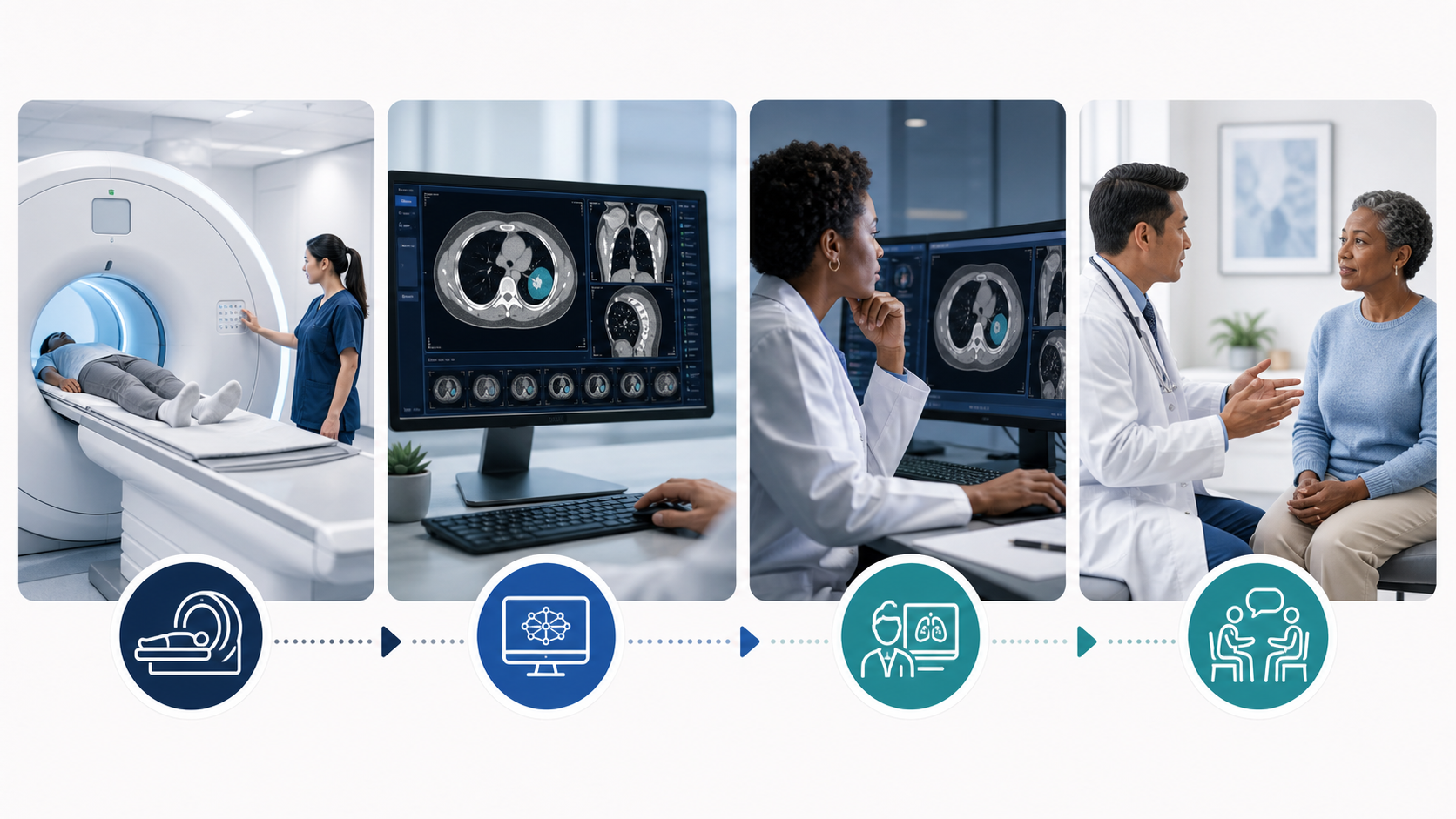
A supervised imaging workflow: the examination is performed, software analyzes the images, a qualified clinician reviews the output, and the findings are considered in the patient's clinical context. Credit: Biomed Atlas editorial illustration.
Image acquisition: A technologist performs the X-ray, CT, MRI, ultrasound or other examination using an appropriate clinical protocol.
AI processing: The system analyzes the images for the particular task it was designed to perform.
Clinical review: A radiologist or other qualified professional reviews the images, AI output and relevant patient information.
Interpretation: The healthcare team decides what the findings mean and whether additional testing, treatment or follow-up is needed.
The safest way to understand imaging AI is as supervised assistance. The software may make one part of the process faster or more consistent, but it does not possess a clinician's complete understanding of the patient.
Where is AI used in medical imaging?
X-rays and chest imaging
AI tools may help identify patterns associated with lung nodules, pneumothorax, pneumonia, fractures or misplaced medical devices. Some systems are designed for worklist prioritization, meaning they can move an examination with a suspected urgent finding closer to the top of a radiologist's queue.
A triage alert is not a final diagnosis, and an examination that is not flagged still requires appropriate review.
CT scans
In CT imaging, AI may support reconstruction, segmentation, automated measurement, dose-related workflow optimization and selected detection tasks. Examples include software used in stroke imaging, pulmonary embolism assessment, internal bleeding, cardiovascular imaging and oncology.
MRI scans
AI may help reconstruct MRI images, reduce noise, outline anatomy and quantify changes. It can also assist with selected neurological, musculoskeletal, cardiac and cancer-imaging tasks.
MRI remains sensitive to acquisition technique, motion and protocol differences, so model performance can change when the operating environment changes.
Mammography and breast imaging
Some systems assist with the detection or characterization of findings on mammography, breast ultrasound or MRI. They may act as an additional reader or draw attention to an area that needs closer inspection.
Screening applications need careful evaluation because both missed cancers and unnecessary recalls can affect patients.
Ultrasound
AI can support image acquisition, anatomical measurements, quality checks and selected interpretation tasks. Because ultrasound depends heavily on how the examination is performed, an algorithm cannot necessarily compensate for incomplete or poor-quality acquisition.
Nuclear medicine and PET
AI may help reconstruct images, segment organs or lesions, quantify tracer uptake and compare findings over time. These measurements can support diagnosis, staging or treatment-response assessment but still need interpretation in the context of the complete examination.
What does the FDA's 2026 AI-device list show about imaging?
Radiology remains the most visible clinical category on the FDA's AI-enabled medical-device list. The list is not a real-time census and the FDA states that it is not comprehensive, but it provides a useful view of where regulated AI is entering clinical practice.
Selected 2026 entries include:
Device | Imaging area | FDA submission | Decision date |
|---|---|---|---|
AiORTA - Plan v2.0 | Radiology | K254207 | Mar 30, 2026 |
Spectral CT Verida Family | CT / Radiology | K253649 | Mar 27, 2026 |
Butterfly Gestational Age Tool | Ultrasound / Radiology | K252148 | Mar 27, 2026 |
Automated Aortic Stenosis Software (AutoAS) | Cardiovascular imaging | K254161 | Mar 27, 2026 |
AI-Rad Companion Brain MR | MRI / Radiology | K253057 | Jan 22, 2026 |
These examples are not a complete list. Regulatory status, intended use and current information should be confirmed in the FDA record for the individual device.
For a fuller explanation of 510(k), De Novo and PMA pathways and selected 2026 devices, see FDA AI-Enabled Medical Devices: 2026 List, Uses and Regulation.
What are the potential benefits of AI in medical imaging?
Faster prioritization of urgent examinations
A system designed for worklist prioritization may alert a radiology team when an examination contains a pattern associated with an urgent condition. If the tool performs reliably and fits the workflow, this can shorten the time before a clinician reviews the case.
It does not guarantee that every urgent finding will be detected.
More consistent measurements
Repeated measurements of lesions or anatomical structures can be time-consuming and may vary between observers. Automated or semi-automated measurements may improve consistency, particularly when a patient is followed over time.
The clinician still needs to confirm that the software measured the correct structure.
Image reconstruction and quality improvement
AI-based reconstruction can reduce noise or help produce useful images from different acquisition strategies. Whether this translates into a shorter examination or reduced radiation exposure depends on the modality, scanner, protocol and specific technology.
AI should therefore not be described as automatically reducing radiation dose.
Support for repetitive workflow
Software can automate selected steps such as labeling anatomy, comparing prior measurements or organizing image series. This may reduce repetitive work, although poorly integrated systems can create additional alerts and checking tasks.
Does AI in medical imaging improve patient outcomes?
Technical accuracy and patient benefit are not the same thing.
An algorithm can perform well on a benchmark or retrospective dataset without proving that its use improves treatment, reduces complications or changes long-term outcomes.
Real clinical value may depend on whether the tool:
reduces the time to review an urgent examination;
improves detection without creating excessive false alarms;
reduces unnecessary repeat imaging;
improves consistency between readers;
changes clinical management appropriately;
reduces avoidable delays;
or improves a meaningful patient outcome.
This is why prospective and real-world evaluation matters. A model's performance should ultimately be judged in the workflow where it is expected to help patients, not only on a curated test dataset.
What does the latest evidence say about trustworthy imaging AI?
In 2026, the research conversation has moved beyond asking whether an algorithm can achieve a high accuracy score.
A systematic review of responsible AI in medical imaging emphasized that trustworthy systems need more than diagnostic accuracy. Important dimensions include transparent reasoning, equitable performance across patient subgroups, privacy protection, calibrated uncertainty and clinical trustworthiness.
The FUTURE-AI framework likewise organizes trustworthy medical AI around six principles: fairness, universality, traceability, usability, robustness and explainability.
Dimension | What clinicians and health systems should ask |
|---|---|
Fairness | Does performance remain acceptable across clinically relevant patient groups? |
External validation | Was the system tested outside the data and institution where it was developed? |
Robustness | Does performance hold across scanners, protocols, image quality and changing clinical conditions? |
Explainability | Does the information shown to the user genuinely help them understand and verify the output? |
Traceability | Can the organization identify the model version, inputs, output and relevant workflow history? |
Monitoring | Is performance checked after deployment and after software or workflow changes? |
Explainability itself should not be treated as a guarantee of correctness. A 2026 perspective in the American Journal of Roentgenology argues that useful explainability needs technical robustness, adaptation to the end user and alignment with the actual clinical task.
What are foundation models in medical imaging?
Most traditional imaging AI systems are developed for a relatively narrow task, such as detecting a particular abnormality or segmenting a structure.
Foundation models take a broader approach. They are trained on large and diverse datasets and can potentially be adapted to several downstream tasks instead of being built for only one purpose.
In August 2026, a Nature Biomedical Engineering perspective described foundation models as part of a shift from task-specific imaging algorithms toward broader backbone models that may combine imaging with pathology, clinical records or genomic information.
The potential is significant, but the same review highlighted a gap between benchmark performance and real clinical value. Representative training data, domain robustness, interpretability, workflow integration and prospective outcome-based validation remain important limitations.
Broad capability should therefore not be confused with broad clinical authorization. A foundation model still needs evaluation for the task and setting in which it will actually be used.
What is multimodal AI in medical imaging?
Multimodal AI combines more than one type of information. In imaging, that could mean combining CT or MRI data with pathology, laboratory results, clinical notes, genomic information or other patient data.
This may help a system consider more of the context that clinicians already use when interpreting a scan. Research published in 2026 describes rapid progress in multimodal biomedical imaging, while continuing to identify data quality, interpretability and ethical issues as important barriers.
Multimodal capability may make AI more useful, but it also increases the amount of information and the number of failure points that need to be validated.
What is agentic AI in radiology?
Agentic AI goes beyond producing one prediction or generated answer. An imaging agent can be designed to work through several steps, such as retrieving prior studies, consulting approved data sources, selecting an analysis tool, comparing findings and preparing information for professional review.
Peer-reviewed radiology literature in 2026 describes agentic AI as a potentially important direction for workflow automation and integration. At the same time, reviews emphasize that much of the evidence is still technical or exploratory and that prospective real-world studies are needed.
This does not mean radiology is moving toward unsupervised autonomous diagnosis. The more actions a system is allowed to perform, the more important permissions, audit trails, validation and human approval become.
For a broader explanation, see Agentic AI in Healthcare: How AI Agents Work, Uses, Risks and Regulation in 2026.
What are the main risks and limitations?
False negatives and false positives
An AI system can miss a genuine abnormality or flag a normal structure as suspicious. A false negative may delay further assessment, while a false positive can contribute to anxiety, extra imaging or unnecessary procedures.
Performance may not transfer to every hospital
A model trained using particular scanners, protocols and patient groups may perform differently elsewhere. Differences in disease prevalence, image quality, demographics and local workflow can change results.
This is why external validation and local performance monitoring matter.
Bias and unequal performance
An overall accuracy figure can hide poorer performance in particular patient groups. Imaging AI should be evaluated across clinically relevant populations rather than assumed to perform uniformly.
Automation bias
Clinicians may place too much confidence in a software suggestion or overlook an abnormality because the system did not flag it. The opposite problem can also occur: too many low-value alerts can make users less responsive to the system.
Limited or misleading explanations
A heatmap, highlighted region or explanation can help a user inspect an AI output, but it does not prove that the prediction is correct. Explainability should support clinical verification rather than become a substitute for validation.
Changes over time
Clinical practice, scanners, software and patient populations change. These shifts can affect model performance even when a system worked well during its original evaluation.
Version control, quality assurance and post-deployment monitoring are therefore part of safe use.
Privacy and cybersecurity
Medical images can contain protected health information and may pass through several connected systems. Organizations need to protect images, metadata, credentials, interfaces and software updates.
Patients should not upload identifiable scans to a public AI service unless they understand how the service stores, processes and uses the information.
Does FDA authorization mean an AI imaging tool is completely accurate?
No. FDA authorization means that a device satisfied the applicable requirements for its particular regulatory pathway and intended use. It does not mean the software is error-free, appropriate for every imaging task or superior to a clinician in every setting.
The FDA list as a whole also contains devices that reached the market through different pathways. Many are cleared through 510(k), some are authorized through De Novo, and some higher-risk devices may be approved through Premarket Approval.
For an individual product, useful questions include:
What exact task and patient population is the device authorized for?
Is it intended for detection, triage, measurement, reconstruction or another purpose?
What scanners and image-acquisition conditions were included in evaluation?
Was it tested at more than one institution?
What are the clinically important false-positive and false-negative consequences?
How is performance monitored after deployment and after software updates?
What happens when the clinician and software disagree?
Will AI replace radiologists?
Current imaging care involves much more than pattern recognition.
Radiologists select and adapt examinations, assess image quality, integrate prior studies and clinical history, communicate urgent findings, discuss uncertainty, consult with other clinicians and help guide additional testing or treatment.
AI is more likely to change individual tasks than eliminate the need for accountable clinical judgment. Some repetitive functions may become increasingly automated, while radiologists may spend more time supervising systems, resolving difficult cases, integrating multimodal information and communicating findings.
How should clinicians evaluate an imaging AI system?
Define the task. What exactly is the system supposed to detect, measure, reconstruct or prioritize?
Review the evidence. Was performance tested on independent data and across multiple sites?
Check the population. Does the validation population resemble the patients who will actually receive care?
Check the equipment and protocols. Were similar scanners, image formats and acquisition methods evaluated?
Examine errors. What happens clinically when the system produces a false positive or false negative?
Assess workflow impact. Does the tool save time, or does it create extra alerts and checking?
Define human oversight. Who reviews the output and what happens when clinician and AI disagree?
Monitor after deployment. Performance should be reassessed as software, scanners and patient populations change.
What should patients ask when AI is used in imaging?
Was AI used to acquire, reconstruct, analyze or prioritize my images?
What specific job did the software perform?
Did a qualified clinician review the complete examination?
Does the AI result affect whether I need another test or treatment?
What happens if the software and clinician disagree?
Is the tool authorized for this particular use?
How is my imaging data protected?
Who should I contact if I have questions about the report?
A patient should not delay urgent medical care while seeking an automated interpretation. New or severe symptoms require assessment by an appropriate healthcare professional.
The bottom line
AI is becoming an important part of medical imaging, but the most useful way to evaluate it is still remarkably practical: What job is this system performing, how good is the evidence for that job, and who is responsible for checking the result?
Medical imaging AI can help with reconstruction, measurements, detection, triage and workflow. Newer foundation, multimodal and agentic systems may eventually connect more parts of the imaging process. Their clinical value will depend on whether performance remains reliable across real patients, scanners and healthcare settings rather than only in research demonstrations.
The goal is not simply to put more AI into radiology. It is to use AI where it makes imaging safer, faster or more informative without weakening professional judgment or patient protection.
Key takeaways
AI can support image acquisition, reconstruction, measurement, segmentation, detection, prioritization and workflow.
Radiology remains one of the most prominent categories on the FDA's AI-enabled medical-device list.
FDA authorization applies to a specific device and intended use; it does not mean the software is flawless or suitable for every patient.
Technical accuracy does not automatically prove improved patient outcomes.
Trustworthy imaging AI requires attention to fairness, external validation, robustness, traceability, usability, explainability and post-deployment monitoring.
Foundation models and multimodal AI are expanding the range of tasks imaging systems may support, but real-world validation remains essential.
Agentic AI is emerging in radiology, but current evidence remains more exploratory than established routine clinical deployment.
False positives, false negatives, bias, automation bias, privacy and performance changes remain important risks.
Qualified clinicians remain responsible for interpreting imaging findings in the context of the individual patient.
Frequently asked questions
What is AI in medical imaging?
AI in medical imaging refers to software designed to perform defined tasks involving medical images, such as reconstruction, segmentation, measurement, detection, triage or workflow support.
Can AI read an X-ray without a radiologist?
Some software can analyze an X-ray for a defined task, but that output is not necessarily a complete interpretation. In routine care, qualified clinicians remain responsible for interpreting examinations and making clinical decisions.
Is AI more accurate than a radiologist?
There is no single answer. Performance depends on the exact task, patient population, disease prevalence, equipment, study design and comparison method. A system that performs well on one narrow task should not be assumed to outperform radiologists across complete examinations.
Can AI detect cancer earlier?
Some AI tools are designed to help detect or characterize suspicious findings in particular screening or diagnostic examinations. Whether they improve earlier detection in practice depends on the tool, population, workflow and supporting clinical evidence.
Does AI reduce radiation exposure?
Some reconstruction or acquisition technologies may support useful images under different protocols, but AI does not automatically reduce radiation. Exposure depends on the examination, scanner settings and clinical protocol.
What is a foundation model in medical imaging?
A foundation model is a broadly trained AI model that can potentially be adapted to several downstream imaging tasks. It may also combine imaging with other clinical information. Broad capability does not remove the need for task-specific validation and appropriate regulatory review.
What is agentic AI in radiology?
Agentic AI refers to systems that can work through several steps toward a defined goal, potentially retrieving information and using approved tools. In radiology, the technology remains emerging and requires careful human oversight, permissions and auditability.
Should I upload my scan to a chatbot?
Uploading identifiable medical images to a general public chatbot may expose sensitive health information and may not provide a reliable diagnostic interpretation. Discuss the official report with the clinician who ordered the examination or a qualified imaging specialist.
How do I know whether AI was used on my scan?
You can ask the imaging facility or clinician whether AI was used for acquisition, reconstruction, measurement, prioritization or interpretation support, and whether a qualified professional reviewed the complete examination.
Related Biomed Atlas guides
Artificial Intelligence in Healthcare: Uses, Benefits, Risks and Examples in 2026
FDA AI-Enabled Medical Devices: 2026 List, Uses and Regulation
Generative AI in Healthcare: Uses, Risks and Regulation in 2026
Agentic AI in Healthcare: How AI Agents Work, Uses, Risks and Regulation in 2026
Sources and evidence
U.S. Food and Drug Administration. Artificial Intelligence-Enabled Medical Devices. Updated periodically.
U.S. Food and Drug Administration. Guiding Principles for Transparency of Machine Learning-Enabled Medical Devices.
Kondylakis H, et al. A Review of Methods for Trustworthy AI in Medical Imaging: The FUTURE-AI Guidelines. IEEE Journal of Biomedical and Health Informatics. 2026.
Savage CH, et al. Explainable Artificial Intelligence for Medical Imaging: A Framework for Bridging the AI Trust Gap. American Journal of Roentgenology. 2026.
Responsible artificial intelligence in medical imaging: a systematic review. 2026.
Muneer A, et al. Foundation models in biomedical imaging: turning hype into reality. Nature Biomedical Engineering. 2026.
Bridging modalities with AI: a review of AI advances in multimodal biomedical imaging. Communications Engineering. 2026.
Chinniah P, et al. Review of agentic artificial intelligence in radiology: from current clinical integration to future innovations. Clinical Radiology. 2026.
Khosravi B, et al. Agentic AI in Radiology: Evolution from Large Language Models to Future Clinical Integration. Radiology: Artificial Intelligence. 2026.
Radiological Society of North America and American College of Radiology. How Artificial Intelligence Is Transforming Medical Imaging.
Centers for Disease Control and Prevention. Radiation in Healthcare: Imaging Procedures.
World Health Organization. Ethics and Governance of Artificial Intelligence for Health: Guidance on Large Multi-Modal Models.
Evidence reviewed: August 21, 2026. Medical-imaging AI, device listings and regulatory guidance can change. Biomed Atlas provides general educational information and does not replace professional medical or regulatory advice.
Medical information notice
Atlas content is intended for education and general information. Seek advice from a qualified healthcare professional for personal symptoms, treatment decisions, or urgent medical concerns.
How this article is maintained
Atlas uses editorial review, evidence checks, visible update dates and a public correction route. Educational content does not replace professional medical advice.
Continue reading
Continue your recent reading
Pick up another medical or biomedical article you opened earlier on this device.
Continue exploring
More medical and biomedical reading
Continue with editorially selected and category-matched articles designed to help readers explore the subject in greater depth.
AI in Drug Discovery: How Artificial Intelligence Is Changing Drug Development in 2026
Artificial intelligence is being used to identify drug targets, screen compounds, design new molecules, predict toxicity and support clinical development. This guide explains how A...
AI Clinical Decision Support: How It Works, Uses, Risks and FDA Regulation in 2026
AI clinical decision support can help clinicians interpret patient information, estimate risk, consider diagnoses and review treatment options. This guide explains how AI-powered C...
Digital Twins in Healthcare: How Virtual Patient Models Work, Uses, Benefits and Risks in 2026
Digital twins are patient-specific computational models that can be updated with real health data and used to simulate possible future outcomes. This guide explains how healthcare...
Agentic AI in Healthcare: How AI Agents Work, Uses, Risks and Regulation in 2026
Agentic AI can go beyond generating answers by planning steps, using tools and carrying out approved actions across healthcare workflows. This guide explains how healthcare AI agen...
AI Medical Scribes: How Ambient AI Works, Benefits, Risks and Privacy in 2026
AI medical scribes can listen during clinical consultations and turn conversations into draft medical notes, letters and other documentation. This guide explains how ambient AI scr...
FDA AI-Enabled Medical Devices: 2026 List, Uses and Regulation
The FDA maintains a growing list of artificial intelligence-enabled medical devices authorized for marketing in the United States. This guide explains what the FDA list means, the...
Questions & Reader Feedback
Help improve this Atlas article
Ask a general question, comment on the article, suggest a correction, or propose a new topic. Nothing is published automatically. Every submission is reviewed by the Atlas editorial team first.
Was this article helpful?
0 helpful · 0 needs improvement